Molecular Imaging

Molecular Breast Imaging Systems

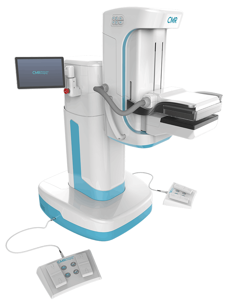
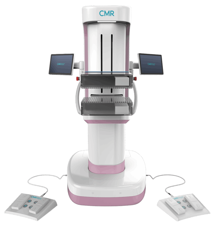
CMR Molecular Breast Imaging

A continuum of information for women's breast health to help clinicians...

Molecular Breast Imaging (MBI) is a type of functional imaging i.e., the pictures it creates show differences in the activity of breast tissue.  Breast tissue that contains cells that are rapidly growing and dividing, such as cancer cells, appears brighter than less active tissue.

Dr. Deborah Rhodes, M.D., a member of the Mayo Clinic Cancer Center, stated, "MBI substantially increases detection rates of invasive cancers in dense breasts without an unacceptable high increases in false positive findings has important implications for breast cancer screening decisions. MBI has a more favorable balance of additional invasive cancers detected versus additional biopsies incurred relative to other supplemental screening options."

The pictures generated during MBI is the result of an injection of a small amount of radioactive tracer.  The tracer attaches to the cancer cells which are detected by the gamma camera of the MBI system.




GettyImages-526640573-59036dcd5f9b5810dc0

LumaGEM® MBI

LumaGEM represents a significant leap forward in technology and performance with fully digital, dual head camera detectors.  With these improvements, LumaGEM customers have been able to identify cancers as small as 5mm in women with dense breast tissue, with greater than 90% sensitivity and specificity.

  •  Clear resolution - Best on the Market
  •  Proven/published lower dose capabilities
  •  High accuracy with Low false negatives
          (high sensitivity, 91%) and Low false
          positives (high sensitivity, 93%)
  •  Patient friendly, comfortable exam with
          minimal compression
  •  Alternative for women contraindicated
          for MRI
CMR-Naviscan LumaGEM

SOLO II™ Breast PET Scanner

SOLO II™ Breast PET Scanner (also called positron emission mammography or PEM) produces high-resolution images with far more detail than conventional breast imaging. Tomographic images of lesions with resolution down to 1.66 mm, about the size of a grain of rice, provide valuable clinical data on invasive and non-invasive diseases across the continuum of care. From initial staging to ongoing post-surgical disease management, the SOLO II provides a metabolic perspective allowing physicians to provide optimal patient outcomes.

  • 3-D Tomographic Imaging and
          automated software
  • Highest sensitivity and
           specificity
  • Precise lesion targeting
  • High 1.6mm spatial resolution
  • SOLO II gently steadies the
          breast without compression


  • Minimal scan times with few to
           no claustrophobic issues
  • Patient can sit comfortably
  • Compact and portable
  • Easy to use
  • MIMviewer image & analysis
          software


CMR Naviscan

CMR X-Ray, LLC News

Designer, manufactuer, installer, and servicer of cutting edge medical imaging systems and supportive equipment.

CMR X-Ray, LLC has demonstrated ability to translate your medical imaging needs into medical diagnostic imaging solutions. Our cutting edge Imaging Generators and the CMR X-Ray patented power assist are designed to work seamlessly with both radiological and molecular imaging systems.

As a certified SDVOSB, CMR X-Ray, LLC can provide our Federal Health Network access to the latest state-of-the-art imaging systems. CMR X-Ray, LLC works with agencies of the U.S. federal government (e.g., The Department of Veteran Affairs or VA) to deliver the highest quality imaging systems to the federal medical community.

Contact Us

GET ANSWER... SHARE INFORMATION

Your questions, ideas, and recommendations are key to CMR X-Ray, LLC

CMR X-Ray LLC

IMAGING INNOVATIONS

CMR X-Ray, LLC